CT Scan Vs MRI: Understanding The Differences, Uses, And Benefits
Have you ever wondered why your doctor might recommend a CT scan over an MRI, or vice versa? When it comes to medical imaging, these two technologies are among the most commonly used diagnostic tools, but they work in fundamentally different ways and serve distinct purposes. Understanding the differences between CT (Computed Tomography) and MRI (Magnetic Resonance Imaging) scans can help you make informed decisions about your healthcare and better understand your diagnosis and treatment options.
What Are CT Scans and MRI Scans?
Both CT scans and MRI scans are sophisticated medical imaging techniques used to capture detailed images of the body's internal structures for diagnosis. While they may seem similar on the surface, each method works differently and has its own unique advantages.
CT scans use X-rays to create cross-sectional images of the body. The machine rotates around the patient, taking multiple X-ray images from different angles, which are then processed by a computer to create detailed slices of the body. This technology is particularly effective at imaging bones, blood vessels, and soft tissues simultaneously.
- You Wont Believe Ice Spices Weight Loss Method Its A Total Scandal
- You Wont Believe What Was Leaked From Walmart St Croix Falls Back Room
- Gypsy Rose And Ryan Andersons Secret Sex Tape Leaked You Wont Believe Whats Inside
MRI scans, on the other hand, use powerful magnetic fields and radio waves to generate detailed images. The magnetic field aligns hydrogen atoms in the body, and radio waves disrupt this alignment. When the radio waves are turned off, the atoms return to their original position, emitting signals that are detected and converted into images by the scanner.
How Do These Imaging Technologies Work?
Understanding the fundamental differences in how CT scans and MRI scans work helps explain why they're used in different situations.
CT Scan Technology
CT technology relies on X-ray beams that pass through the body from multiple angles. As the X-rays travel through different tissues, they're absorbed at different rates depending on the density of the material they encounter. Dense structures like bones absorb more X-rays and appear white on the images, while less dense tissues like muscles and organs appear in shades of gray, and air-filled spaces appear black.
- Snoqualmie Pass Roads Exposed Nude And Defenseless The Horrific Truth Unfolds
- From The Vault The Viral Leak Exposing A Scandal That Changed History
- The Viral Louisville Mens Basketball Twitter Disaster Sex Lies And Leaked Dms That Destroyed The Team
The computer processes the X-ray data to create cross-sectional images, or "slices," of the body. These slices can be viewed individually or stacked together to create a three-dimensional representation of the area being examined.
MRI Technology
MRI technology is more complex and relies on the behavior of hydrogen atoms in the body. Since the human body is composed largely of water, which contains hydrogen atoms, MRI can create detailed images of virtually any part of the body.
The powerful magnetic field in an MRI machine aligns the protons (hydrogen atoms) in the body. Radiofrequency pulses are then applied, temporarily knocking these protons out of alignment. When the radiofrequency pulse is turned off, the protons realign with the magnetic field, releasing energy that's detected by the scanner. The time it takes for different tissues to realign and the amount of energy released varies, allowing the computer to differentiate between various tissue types.
Which Imaging Test Do You Need?
Your healthcare provider chooses between a CT scan and an MRI based on several factors, including the specific condition being investigated, the part of the body being examined, and your individual medical circumstances.
Common Uses for CT Scans
CT scans are particularly valuable for:
- Detecting bone fractures and joint problems
- Identifying internal bleeding and injuries from trauma
- Diagnosing and staging cancer
- Evaluating lung and chest problems
- Guiding procedures such as biopsies or surgeries
- Detecting blood clots and vascular conditions
CT scans are often the first choice for emergency situations because they're faster and can quickly identify life-threatening conditions like internal bleeding or stroke.
Common Uses for MRI Scans
MRI scans excel at imaging soft tissues and are particularly useful for:
- Evaluating brain and spinal cord abnormalities
- Diagnosing joint and soft tissue problems
- Detecting tumors and their characteristics
- Assessing heart and blood vessel structures
- Evaluating pelvic organs and reproductive system issues
- Diagnosing multiple sclerosis and other neurological conditions
MRIs create detailed images of soft tissues like nerves and ligaments that CT scans cannot visualize as clearly, making them invaluable for neurological and musculoskeletal conditions.
Comparing Safety, Comfort, and Cost
When considering which imaging test is right for you, it's important to understand the differences in safety, comfort, and cost between CT scans and MRI scans.
Safety Considerations
CT scans involve exposure to ionizing radiation, which is a consideration for certain patients, particularly pregnant women and those who require frequent imaging. However, the radiation dose from a typical CT scan is relatively low, and the benefits usually outweigh the risks for most diagnostic situations.
MRI scans do not use ionizing radiation, making them safer for patients who need repeated imaging or who cannot be exposed to radiation. However, the strong magnetic field can be dangerous for patients with certain metal implants, pacemakers, or other metallic devices.
Comfort and Patient Experience
The patient experience differs significantly between the two technologies:
CT scans are generally quick and painless, typically taking only a few minutes to complete. The machine is open and less confining than an MRI, which can be beneficial for patients with claustrophobia.
MRI scans take longer, often 30-60 minutes or more, and require the patient to remain completely still in a confined space. The machine produces loud knocking and buzzing noises, which can be disturbing to some patients. However, many facilities provide earplugs or headphones to help with the noise.
Cost Differences
Generally, CT scans are less expensive than MRI scans. The equipment for CT scanning is less costly to purchase and maintain, and the shorter scan times mean facilities can accommodate more patients. However, costs vary significantly depending on your location, insurance coverage, and the specific facility.
Special Applications and Advanced Imaging
Beyond standard CT and MRI scans, there are specialized applications and advanced imaging techniques that provide even more detailed information for specific conditions.
PET Scans and Nuclear Medicine
Positron emission tomography, also called PET imaging or a PET scan, is a type of nuclear medicine imaging that uses small amounts of radioactive material called radiopharmaceuticals or radiotracers. These tracers are injected into the body and accumulate in areas of high metabolic activity, such as cancer cells.
Nuclear medicine uses small amounts of radioactive material to diagnose, evaluate, and treat various diseases. PET scans are often combined with CT scans (PET/CT) to provide both functional and anatomical information, making them particularly valuable for cancer diagnosis and treatment monitoring.
Specialized Applications
MRI ultrasound is becoming increasingly popular for certain conditions, particularly groin hernias. In one hospital study of 215 patients, 70% had ultrasound before surgery compared to 28% who had CT. If your hernia tends to come and go, mentioning that to the technologist beforehand can help them adjust the technique to capture the condition when it's visible.
For facial nerve palsy, the choice between imaging modalities depends on the suspected cause. While MRI is excellent for evaluating soft tissue structures and can detect inflammation or compression of the facial nerve, CT may be preferred if bone involvement is suspected.
The Role of Imaging in Cancer Diagnosis and Treatment
Both CT scans and MRI scans play crucial roles in cancer diagnosis and treatment, often working together to provide comprehensive information.
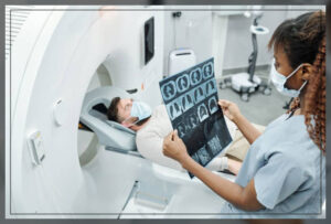
A CT scan, shown here, and MRI are both commonly used in diagnosing and staging cancer. CT scans are particularly valuable for detecting the size and location of tumors, evaluating whether cancer has spread to lymph nodes or other organs, and monitoring treatment response. They're especially useful for cancers in the chest, abdomen, and pelvis.
MRI scans excel at characterizing tumors, particularly in the brain, spinal cord, and soft tissues. They can help distinguish between benign and malignant tumors and provide detailed information about tumor boundaries, which is crucial for surgical planning.
Advanced Heart Imaging
A coronary artery calcium (CAC) test is a kind of heart scan that uses CT technology to detect calcium deposits in the coronary arteries, which can indicate the presence of coronary artery disease. This non-invasive test helps assess heart disease risk and can guide preventive treatment decisions.
Cardiac MRI provides detailed images of heart structure and function, making it valuable for evaluating heart muscle damage, congenital heart defects, and other cardiac conditions that might not be visible on other imaging tests.
Limitations and Considerations
While both CT and MRI scans are powerful diagnostic tools, they have limitations and aren't appropriate for every situation.
Proving a brain injury that is not visible on an MRI or CT scan requires a deep understanding of how these injuries work and how medical professionals view "invisible" disabilities. Standard scans miss brain injuries that don't cause structural damage, such as mild traumatic brain injuries or concussions.
Why standard scans miss brain injuries: CT scans and MRIs are designed to look for structural damage, like skull fractures or bleeding. They may not detect functional changes, microscopic damage, or chemical imbalances that can occur with certain types of brain injuries.
Emerging Technologies and Open Science
The field of medical imaging continues to evolve with new technologies and approaches to data sharing and research.
OpenNeuro is a free platform for sharing, browsing, and managing neuroimaging data, fostering open and reproducible research in the field. This initiative helps advance our understanding of brain function and disease by making neuroimaging data accessible to researchers worldwide.
Choosing the Right Imaging Test for Sinus and Head Conditions
When it comes to sinus and head conditions, the choice between imaging modalities depends on the specific symptoms and suspected conditions.
Get the CT scan sinus facts on what they see: CT scans are excellent for evaluating sinus anatomy, detecting inflammation, polyps, and structural abnormalities. They provide detailed images of bone structures and can identify blockages or anatomical variations that might contribute to chronic sinusitis.
Understand the powerful difference between sinus imaging and brain scans for your safety and care. While a CT scan of the sinuses focuses on the nasal passages and surrounding structures, a CT scan of the brain reveals detailed images of brain structures, detecting bleeding, tumors, fractures, and other abnormalities quickly and accurately.
Conclusion
Understanding the differences between CT scans and MRI scans empowers you to be an informed participant in your healthcare journey. Both technologies offer unique advantages and are chosen based on the specific clinical question, the part of the body being examined, and your individual medical circumstances.
CT scans excel at quickly imaging bones, detecting internal bleeding, and providing comprehensive views of the chest and abdomen. They're often the first choice in emergency situations and for cancer staging.
MRI scans provide superior soft tissue contrast, making them ideal for evaluating the brain, spinal cord, joints, and other areas where detailed soft tissue imaging is crucial. They're particularly valuable for neurological conditions, musculoskeletal problems, and characterizing tumors.
The key is working with your healthcare provider to determine which imaging test is most appropriate for your specific situation. Factors such as the suspected condition, the need for speed versus detail, safety considerations, and cost all play a role in this decision.
As medical imaging technology continues to advance, both CT and MRI scans will likely become even more sophisticated, providing clearer images, faster scan times, and more precise diagnostic information. Whether you're facing a routine diagnostic test or dealing with a complex medical condition, understanding these imaging technologies helps you navigate your healthcare with confidence and clarity.